Information for veterinarians
- Information for veterinarians
- Indications
- Rehabilitation and fitness training
- Case Report
- ESVN Paris 2013
- ESVN-ECVN Symposium 2014 - Madrid
- Glossary
Orthomanual veterinary medicine
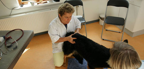
The idea behind orthomanual veterinary medicine is that many cases of lameness and neurological deficiencies are caused by a misalignment of the vertebrae and joints. Orthomanual manipulation is done by applying direct pressure or force on an osseous structure of the spine with a malposition or malfunction, using the hands or a short lever within a segment.
When is orthomanual therapy used?
- Orthopaedic patients
- Lame animals, young and old, acute or chronic
- Animals with arthrosis and degenerative joint disease (DJD)
- Neurological patients
- Animals with intervertebral disc disease (IVDD)
- Animals with weakness in the fore or hind limbs
- Patients with pain in the neck, back or lower back
- Patients with lumbosacral stenosis or instability (DLSS)
- Animals with behavioural problems like restlessness, anxiety and aggression (possibly related to pain)
- Working and sporting dogs (preventive orthomanual sport check-ups)
- Puppies (preventive and educational)
- Senior pets (preventive and educational)
- Conservative (rest, medications)
- Surgical
- Manipulation (manually adjusting misalignments)
Ideally, an orthomanual veterinarian works closely with other disciplines, including diagnostic clinics, neurologists, neurosurgeons and orthopaedic surgeons. Decisions on how to treat a patient are based on thorough clinical, neurological and orthopaedic examination and patient assessment.
Three basic treatment options are available:
A prompt referral is preferred.
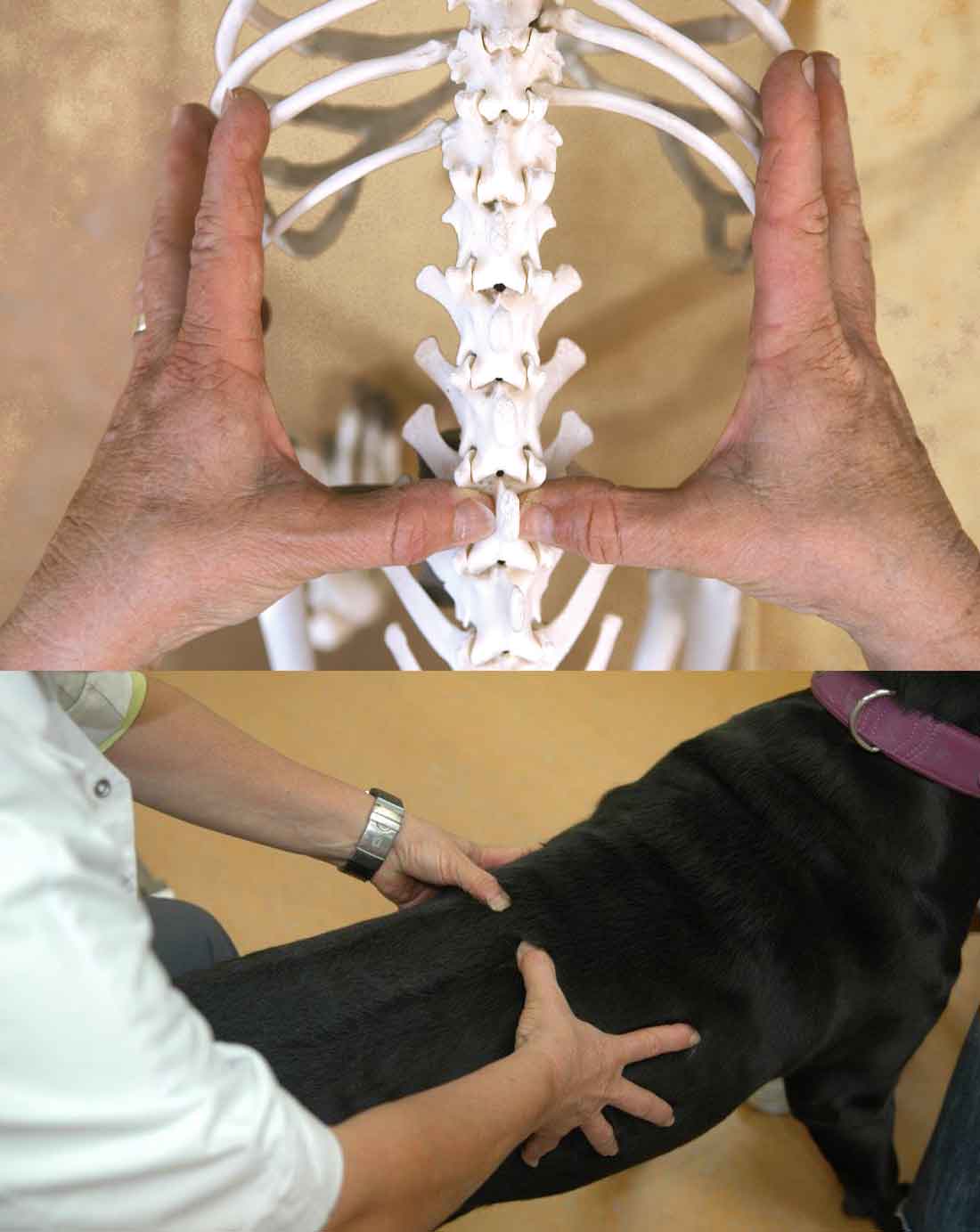
Misaligned vertebrae
Instability of the spine due to, for example, intervertebral disc degeneration, congenital disorder or trauma, can lead to rotation, dislocation or translation of one or more vertebrae.
From the orthomanual perspective, a vertebral position is considered misaligned when its position differs from the longitudinal, ventrodorsal or lateral (dextrosinistral) body axis. Moreover, a vertebral position is considered misaligned when the transverse processes are not parallel. Misalignment of skeletal components can cause loss of function, movement limitations and pain. The aim of the Aharon Method is to adjust misaligned vertebrae and joints, thereby restoring them to a more optimal position and function.
An example
In animals, instability of the low back and misalignment of vertebrae in the neck (lumbosacral instability and Wobbler syndrome, respectively) are all too common. Wobbler syndrome, for instance, is a neurological problem caused by, among other things, malformed vertebrae. This abnormality is well documented in older Doberman Pinschers and younger Great Danes.
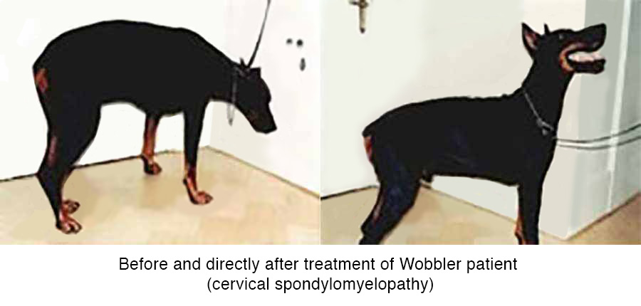
Before and directly after treatment of Wobbler patient (cervical spondylomyelopathy)
Caudal cervical spondylomyelopathy (CCSM)
In CCM, neurological symptoms are caused by compression of the cervical spinal cord. This disease most commonly affects large and giant dog breeds, especially the Great Dane and Doberman Pinscher. The etiology of CCSM is multifactorial and includes vertebral instability, stenosis and/or malformation, hernia and degenerative changes in the vertebral facet joints and ligaments. One or a combination of these leads to progressive stenosis and compression of the cervical spine. The compression may be dynamic, meaning the degree of compression varies with the position of the neck.
Symptoms can occur in very young animals, but may also develop in later life. Typical CCSM signs are wide-based pelvic limb ataxia and pseudohypermetria of the thoracic limbs, or unilateral thoracic limb lameness due to nerve root compression (root signature). Treatment options are conservative or surgical. Prognosis is always guarded and recurrence of symptoms is common.
Orthomanual treatment aims to create more intervertebral space and to stabilize the spine. After treatment, strict rest should be maintained for two weeks. Further, ergonomics-related advice is provided to the owner to better manage the animal’s environment.
Hernia Nucleus Pulposus (HNP)
HNP is an outdated and essentially incorrect term. In veterinary medicine, pathologies of the intervertebral disc are currently referred to as intervertebral disc disease (IVDD). Furthermore, the term cervical disc disease (CDD) is used for lesions in the cervical region, and thoracolumbar disc disease (TLDD) for lesions in the thoracolumbar region. The animal’s neurological state based on the neurological examination is classified as follows:
| Grade | Neurological state | Proprioception | Bladder control | Deep pain perception |
|---|---|---|---|---|
| I | Pain | + | + | + |
| II | Paraparesis | - | + | + |
| III | Paraparalysis | - | + | + |
| IV | Paraparalysis | - | - | + |
| V | Paraparalysis | - | - | - |
In orthomanual veterinary medicine, degeneration of the discs is understood to result in instability of the vertebrae. This instability can cause misalignment of adjacent vertebrae, leading to disc degeneration and, in turn, extrusion or protrusion of the nucleus pulposus or annulus fibrosis. The pressure on the spinal cord, or trauma induced by the kinetic energy component of the extruded disc material, can cause pain and neurological dysfunction.
The orthomanual approach seeks to correct the vertebral misalignment, to reduce the pressure on the intervertebral disc and create an environment that facilitates improvement of the neurological state. Full recovery is defined as neurological grade 0, with a completely normal gait and proprioception and without residual paresis. Clinical recovery with some residual motor deficiency (paresis and/or delayed proprioceptive reflexes) is defined as recovery to grade I.
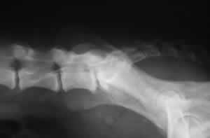
Spondylosis deformans
Spondylosis is a non-inflammatory process associated with degeneration of the annulus fibrosis of the intervertebral disc, in which bony spurs or bridges form between adjacent vertebrae. Spondylosis is known to give few complaints (sometimes stiffness). A distinction must be made between spondylosis and DISH (diffuse idiopathic skeletal hyperostosis). The latter disease causes certain ligaments and tendons to ossify, which affects both the spine and the limbs.
DISH is best known in humans. But dogs, especially Boxers, seem to be susceptible. In dogs, DISH can occur in combination with spondylosis, but the symptoms of DISH are severe (pain, neurological deficit, fractures), in contrast to spondylosis.
The orthomanual approach views the bridging as the body’s attempt to compensate for vertebral instability and therefore as a signal of possible problems in the back. Spondylosis and DISH have also been associated with adjacent segment disease, because the fusion of two vertebrae causes a relative instability of the adjacent vertebrae.
Lumbosacral instability, lumbosacral stenosis: before treatment

Lumbosacral instability, lumbosacral stenosis: a year after treatment
Adjacent segment disease
Pathologies of adjacent areas of the spine (caudally and cranially) after spinal surgery are well known in dogs with cervical spondylomyelopathy. It is a major long-term complication after spinal surgery.
Unlike spinal surgery (for TLDD, CDD, etc.) which produces only local decompression, orthomanual treatment seeks to correct all vertebral misalignments, also addressing areas where subclinical intervertebral disc disease might be present. This results in “decompression” where appropriate, thus preventing adjacent segment disease.
Notes
- Van de Veen et al.; Variance in Manual Treatment of Nonspecific Low Back Pain between Orthomanual Physicians, Manual Therapists, and Chiropractors, Journal of Manipulative and Physiological Therapeutics, 2005; vol. 28.
- Oliver and Lorenz. "Handbook of Veterinary Neurology" 2nd ed. W.B. Saunders, 1993: pages 180-185.
- Morgan J: "Spondylosis deformans in the dog". Acta Orthopaedica Scandinavica Supplementum 1967; vol 96: blz 1-88.